The ankle joint is one of the single stance.
Anatomy and biomechanics
First principle: stability.
It is due to a surface tibiotalar ultra congruent.
Tenon is the talus bone of the “take” in the bi-ankle mortise. The mortise is formed of the distal end of the fibula laterally, and medially by the distal epiphysis of the tibia.
Second principle: the semi mobility.
It is semi mobile with intra-articular mobility in different angular sectors (flexion / extension and varus / valgus).
The elements of the tibiotalar stability and tibiofibular are:
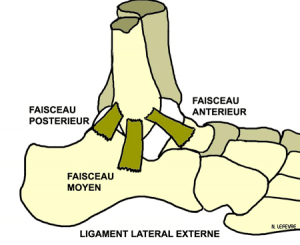
- The complex of the external lateral ligament: anterior tibiofibular ligament, the fibular ligament-calnéo and tibiofibular ligament
- The medial collateral ligament
- The tibiofibular syndesmosis
1 / The complex anatomical of the tibiofibular syndesmosis lower
The syndesmosis is a semi-movable joint between two bones, ligaments or united by a membrane. The tibiofibular joint deviates in dorsiflexion, plantar flexion tightens. There is thus a stress distribution on the slope during the flexion extension tibiotalar.
The ligaments of the tibiofibular syndesmosis are:
- the anterior tibiofibular ligament: it fits the Tillaux tuber to the front edge of the fibular malleolus and provides 36% of the previous congruence biomechanically
- the tibiofibular ligament posterior: it extends the posterolateral tibial tubercle of the posterior side of the fibular malleolus and it is 42% in support of the constraints of the tibiofibular
- the interosseous tibiofibular ligament extended by the interosseous membrane and that it ensures the remaining 22% resistance of the tibiofibular syndesmosis.
There is a mechanical bio Association 2 tibiotalar joints and tibiofibular.
2 / The mechanism of injury of lower tibiofibular
During plantar flexion there is an internal rotation of 30 °, asymmetry Italian cheeks in their orientation and width. Thus the lateral plays converges forward and off, while the medial cheek is more sagittal. The posterior part of the slope is wider than its front portion. The gap in flexion varies so. The anterior tibiofibular ligament hinge made ??during bending tibiotalar extension.
Dorsiflexion tibiotalar, 4 steps to succeed. The lateral malleolus deviates rises slightly and undergoes internal rotation on itself, the posterior tibiofibular ligament is stretched.
Plantar tibiotalar flexion, lateral malleolus is close by contraction of the tibialis posterior, lateral malleolus lowers slightly and then commits an external rotation on itself.The anterior tibiofibular ligament is stretched.
In plantar flexion, tibiotalar joint is less congruent, and constraints are supported by the tibiofibular ligaments.
So there is in plantar flexion with a slope that is wider in front than behind, a tibiofibular mobility increases, a reduction of the articular congruence. The mechanism of injury in forced plantar flexion therefore generates a lesion of the anterior tibiofibular ligament.
During the lateral rotation there is a distraction from the lateral malleolus, a tibiofibular gap increased and therefore a distention of the anterior and posterior tibiofibular ligaments.
During adduction pronation which causes lesions equivalent of a bi-malleolar fracture with fracture of the medial malleolus and syndesmosis sprain. Mechanical forces pass through the medial malleolus by a horizontal line and pass in the external ankle mortise by a vertical line through the tibiofibular ligament and may end with an above-tubercular fibula fracture.This is a lesion adducted forced pronation.
3 / The lesion diagnosis of the tibiofibular syndesmosis
Diagnosing a lesion of the syndesmosis includes an analysis of the mechanism of injury. It is often a hyper plantarflexion associated with rotation. There are serious things to look for, the total impotence, lameness, deformation, crack sensation and not active mobility.
On clinical examination, it feels different bony seats, fibula, tibia, the epiphysis and diaphysis, increasingly getting closer to the pain. It is an elective pain anterolateral feature.
There is also an earlier slurred ankle, the anterolateral pain disappears at the landfill. There are three specific tests: Pain in dorsal flexion and adduction, the pain forced into external rotation and pain reproduced by the sign says Hopkinson or squeeze test. It has a proximal compression calf tibia and fibula causing distal distance and was pain on the syndesmosis.
Additional tests that are carried out to confirm the diagnosis are:
Radiographs are essential. They allow a study of face, profile and internal rotation of the ankle. Also, do not forget to look for a lesion on the entire fibula with radios face and leg profile.Search type of bone injury: ankle fracture, fibular or calcaneus. or bone avulsion on the inner and outer ligament attachment of the ankle.
There is also a search osteochondral lesion of the talus.We seek an indirect sign of ligament injury especially with tibiofibular diastasis lower. Indeed, the gap between the anterior tubercle, posterior of the distal epiphysis of the tibia and fibula are characteristic. A difference of more than 5 mm internal tibiotalar is also characteristic of a severe sprain. It also directs ultrasound which should not be isolated but further because it is an interesting dynamic diagnosis.
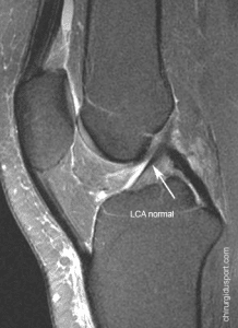
MRI is systematic: the exam eliminates bone injury, diagnose the rupture of the anterior tibiofibular ligament, eliminate posterior tibiofibular injury, eliminate posterior tibiofibular injury to graduate achieving the inter bone membrane lesion. It also uses the classification Kouvalchouk sprains in 2000 into three stages:
- Stage 1: ligament injury without rupture
- Stage 2: ligament rupture without displacement
- Stage 3: tibiofibular diastasis.
4 / The treatment of tibiofibular injury
Treatments vary depending on the classification and accurate lesion diagnosis made ??during the interview with the doctor:
Functional treatment: early rehabilitation without immobilization
Orthopedic treatment: immobilisation by plaster or resin boot for 4-6 weeks because it offers either a functional treatment in stage 1 of a tibiofibular reached
The surgical treatment consists in reducing the fracture displacement or gap between the bone by various mechanisms, a son traction system or a screw or plate system.
Complications of the syndesmosis injury is severe, with sequelae type of pain and instability, and may be associated frequently with osteochondral lesions.
In conclusion, the ankle-sprain is a different éèfibulaire sprain sprain “usual” banal ankle.
It must be sought by the doctor’s clinical examination of the sport, and confirmed by MRI.
The prognosis is related mainly to taking initial therapeutic. You should know that the recovery is long, between 2 and 3 months and in case of inadequate treatment, may expose themselves to sequels.
Doctor Yoann BOHU, Doctor Serge HERMAN, Doctor Nicolas LEFEVRE. – 23 octobre 2013.