Introduction
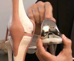
The aim of this operation is to achieve anatomic ACL reconstruction using autologous (patient’s tendon) under arthroscopic control. The principle of TLS is to use a single hamstring tendon graft in short.
Anatomy:
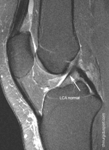
ACL
The anterior cruciate ligament is a short ligament, very durable. It is stretched between the femur and tibia. He participates in the central pivot with the posterior cruciate ligament is behind him. It works in synergy with the external and internal lateral ligament
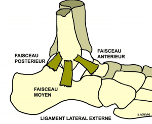
Medial collateral ligament ( MCL ) is the ligament of the inside of the knee. It is long, wide and spread out. It is stretched between the femur and tibia at the top to bottom. It allows the internal stabilization of the knee.
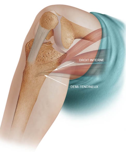
The ACL graft: autologous tendon in half.
The half tendon tendon is one of two hamstring tendons (hamstring). It is end (3 to 4 mm in diameter) and long (about 25 cm)
It is the termination of the semitendinosus that ends on the bridle.
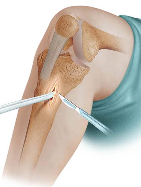
Prélèvement graft
For a short incision almosthorizontal, from 2 cm to thesurface of the tibia, the tendon is removed only half tendon over their entire length with a “stripper”
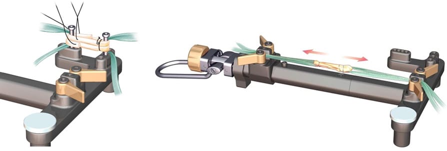
He bent over backwards to get an ACL graft 4 beams or 4 strands whose average diameter is 7 to 9 mm. It’s a short graft 50mm medium length.
At both ends of the graft are passed two strips for fixing the graft in tunnels.
A traction table is used to make a claim to the transplant
Arthroscopic time
The procedure is performed arthroscopically.
Two small incisions of 5 mm on either side of the ball will allow to move the camera and instruments to perform the ligament
The first operative step is the exploration of the knee:
essential step of the surgery, it allows for a full assessment of the lesions of the knee noble elements (menisci, cartilage, ligaments other …)
Treated if necessary these peripheral lesions then realizes ligamentoplasty
The preparation of the notch allows the cleaning of the axial face of the lateral condyle and visualize the area well “over the top” in order to delimit the dial 9-12 and 12-15h for a right knee to the left knee.
Referred femoral
The femoral tunnel is drilled under arthroscopic control inwards with a femoral viewfinder (FH). With the knee at 90 ° flexion. Tunneling on the spindle 4.5mm diameter is performed using the drill 4,5mm.
The femoral stall is then excavated using an auger to specific fins. Different diameters are provided to dig a little cell corresponding to the graft diameter measured at each end.
The arthroscope is controlling the retrograde manual digging.
Then a TLS spindle guide cannula is positioned, being screwed into the threaded cavity in order to maintain the correct axis of the tunnel.
Tibial referred
The tibial tunnel is drilled under arthroscopic control inwards with a tibial viewfinder (FH). With the knee at 90 ° flexion. Tunneling on the spindle 4.5mm diameter is performed using the drill 4,5mm.
The tibial stall is then excavated using an auger to specific fins. Different diameters are provided to dig a little cell corresponding to the graft diameter measured at each end.
The arthroscope is controlling the retrograde manual digging.
Then a TLS spindle guide cannula is positioned, being screwed into the threaded cavity in order to maintain the correct axis of the tunnel.
Passage of son
Tractors son went inwards in the femoral and tibial tunnels using a pass over TLS and recovered by the instrumental track with a prehensile claw.
Retrieving strips
The strips corresponding to the end of the graft for the femur are passed through the loop formed by the towing wire.
The graft is then towed on his bandages and is positioned automatically in the femoral stall. The strips corresponding to the tibial end of the graft are passed through the loop formed by the tractor to tow wire which is in turn and provides a tibial positioning.
Fixing
Placing tls® screw
We begin with the femoral and tibial locking.
Hip screw
The guide pin is positioned between the two femoral strips to feel the touch of the tip of the pin on the graft.
The hip screw 20mm is in place and screw being careful to stop flush with the bone.
Tibial screws
The tibial screw 25mm is put in place and screwed knee flexed to 45 °. The good voltage control graft can be done to hook a new arthroscopic view.
The strips are then cut flush screws.
The procedure finishes with the skin closure leaving, depending on the habits, intraarticular suction drain and / or another in the sampling zone.
Post-operative care
The rehabilitation mobility business from the day after the procedure without any particular limitation. If the procedure took place without technical complications, the TLS system by mounting the mechanical performance are used to allow safe immediate resumption of full support on the operated limb, according to the patient’s abilities.
Doctor Nicolas LEFEVRE, Doctor Yoann BOHU, Doctor Serge HERMAN. – 3 janvier 2015.